Bing Ads
Home » ACLS Course » Acute Coronary Syndrome
Acls-course-img-eng
Acls-course-mob-img

ACLS Course

Our online ACLS certification course includes understanding the physiology of the heart, an overview of BLS and ACLS, ACLS Pharmacology, and AED training. The course is best suited for healthcare/medical professionals. It has 2 years validity. A group of 5 or more can take Read More advantage of group discounts. Read Less
(111,773 reviews with 4.8 stars)

$119.00 $169.00

ChaptersCE CreditsValidityCostDurationECCExam AttemptsWallet Card
174.0 – 8.02 Years$119.003-4 HrsCompliantUnlimitedDownload/Print/Mail
  • Chapters17
  • CE Credits4.0 – 8.0
  • Validity2 Years
  • Cost$119.00
  • Duration3-4 Hrs
  • ECCCompliant
  • Exam AttemptsUnlimited
  • Wallet CardDownload/Print/Mail

Select Course

Chapter 13 : Acute Coronary Syndrome

Acute coronary syndrome or ACS is defined as a sudden, reduced blood flow to the heart. ACS is seen in ST-segment elevation myocardial infarction (STEMI), non-ST-segment elevation myocardial infarction (NSTEMI) or unstable angina. Myocardial ischemia occurs when there is not enough blood supply to the heart muscle and that results from coronary artery disease (CAD). ACS is usually caused by atherosclerosis and coronary thrombosis, but can also be associated with cocaine use. Other conditions that cause ACS include:

  • Unstable Angina (UA)
  • Microemboli
  • Occlusive Thrombus
  • Unstable Plaque
The symptoms associated with ACS include:
  • Chest pain radiating to the left arm and left angle of jaw
  • Shortness of breath
  • Nausea
  • Vomiting
  • Diaphoresis
  • Palpitations
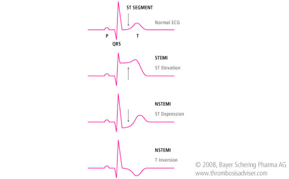
ST-Segment Elevation Myocardial Infarction (STEMI) is an elevation > 1mm in 2 or more contiguous or greater than 2 mm in leads V2 and V3. Unstable Angina (UA) or Non ST-Elevation Myocardial Infarction (NSTEMI) is a depression ≥0.5 mm or greater T-wave inversion. Low Risk Unstable Angina (UA) is an ST segment deviation of <0.5 mm or T wave inversion of 2 mm. Patients may also have normal ECGs. chapter10-1 Scenario:  A 70-year patient comes to the hospital and is complaining of chest pain radiating to his arm and he seems very anxious and out of breath.  Assessment:
  1. Identify signs and symptoms of ischemia or infarction
  2. Monitor vital signs and cardiac rhythm
  3. Be prepared to do CPR and use the AED
  4. IV access
  5. Check cardiac markers, electrolytes and coagulation status
Interventions:
  1. Give aspirin (162 – 325 mg), oxygen if O2 saturation is <90% start oxygen at 4L/min, nitroglycerin sublingual every 3 to 5 minutes, and morphine for pain. Exercise caution when utilizing nitroglycerin.
    1. Be mindful of inferior wall myocardial infarction (MI) and right ventricular (RV) infarction.
    2. Monitor for systolic blood pressure (SBP) below 90mmHg and significant bradycardia (heart rate less then 50/min), or marked tachycardis.
    3. Caution with phosphodiesterase inhibitor usage due to the potential for severe hypotension.
  2. Conduct  a 12-lead ECG and diagnose
Management:
  1. If STEMI on ECG perform rapid fibrinolytic therapy (“clot buster”) or direct reperfusion
  2. If time of onset of symptoms is ≤12 hours – reperfusion:
    1. Door to balloon inflation (PCI) – 90 minutes
    2. Door to needle fibrinolysis – 30 minutes
  3. If time of onset of symptoms is >12 hours, then invasive treatment is recommended
  4. If high risk UA or NSTEMI on ECG check troponin level and start invasive treatment
  5. Immediate medications are
    1. Aspirin
    2. Nitroglycerin
    3. Morphine
    4. Consideration of p2y12 inhibitors (antiplatelet)
  6. Monitor and continue heparin and may add ACE inhibitor and HMG CoA reductase inhibitors if needed
  7. If low risk ACS check cardiac markers, repeat ECG, conduct non-invasive diagnostic test
  8. If no ischemia or infarction discharge and schedule a follow-up
Treatments which are used in ACLS algorithm for ACS are:
  • Oxygen
  • Aspirin
  • Nitroglycerin
  • Morphine
  • Fibrinolytic (thrombolytic) therapy
  • Heparin

The following is an algorithm that shows management of ACS in detail:

acute-coronary-img-01
acute-coronary-img-02