A course in Advanced Cardiovascular Life Support (ACLS) teaches students all about how the heart works and what to do during a cardiac issue. More specifically, ACLS trains healthcare workers to recognise and intervene in cardiac dysrhythmias, cardiopulmonary arrest, stroke, and acute coronary syndrome, among other conditions.
The goal of ACLS training is to improve the survival rate of adults who are experiencing cardiac or neurological crises. At the core of mastering ACLS is the need for a fundamental and in-depth understanding of how the heart works. Read on to find out all about how the heart works.
How Does the Heart Work?
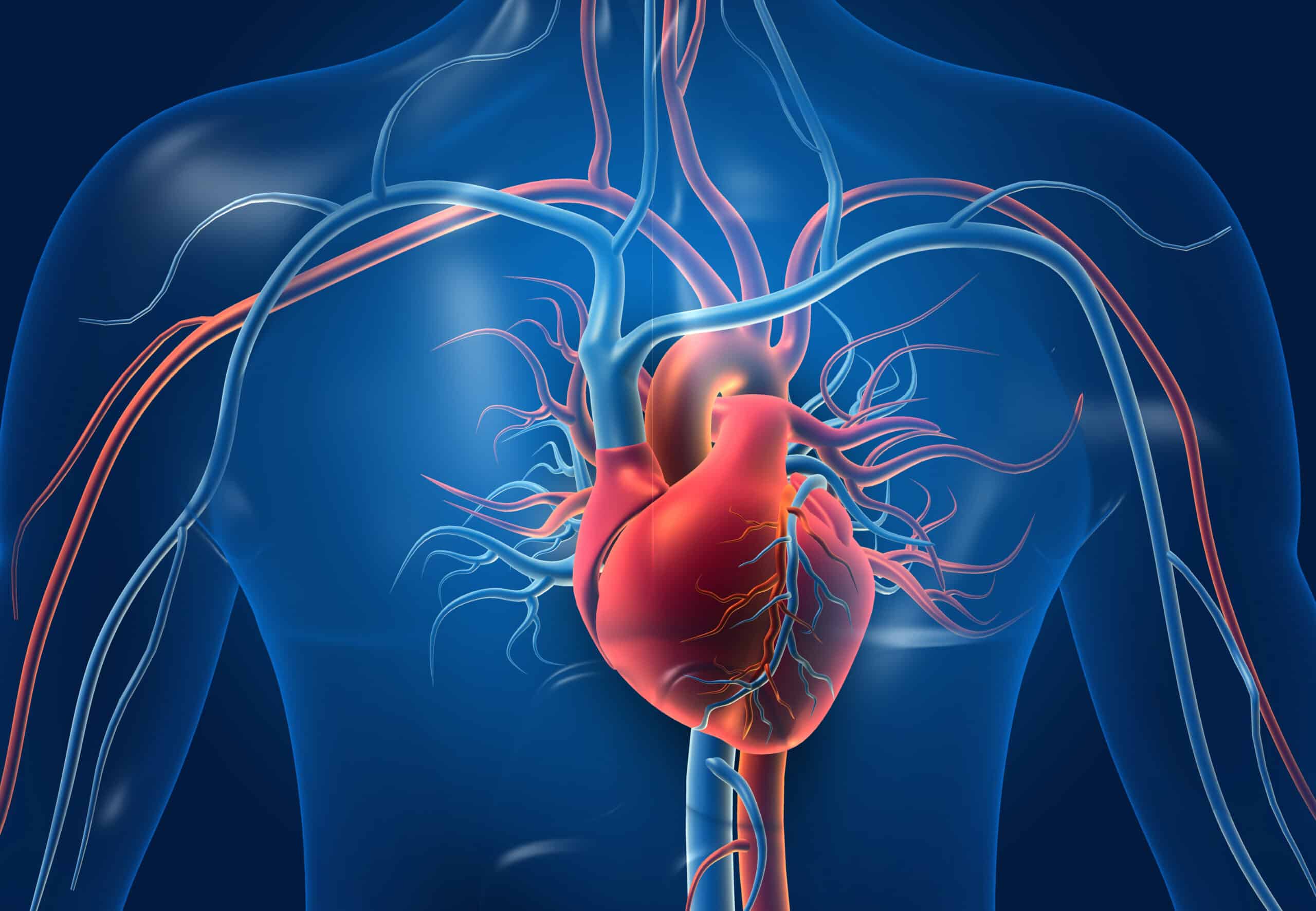
The heart is one of the most vital and hardest working organs of the human body. It is a pumping muscle that is the main element of the cardiovascular system and it is around the size of a fist.
While it has been idealized by poets, writers, and lovers, the heart has a fundamental and necessary purpose. Specifically, it serves to pump oxygenated blood to the body and deoxygenated blood to the lungs, among other things.
The heart beats continuously, and during infancy, the heart beats approximately 120 times per minute. As a person grows, the number of beats per minute in your heart drops to an average of 72 beats per minute.
Heart Anatomy
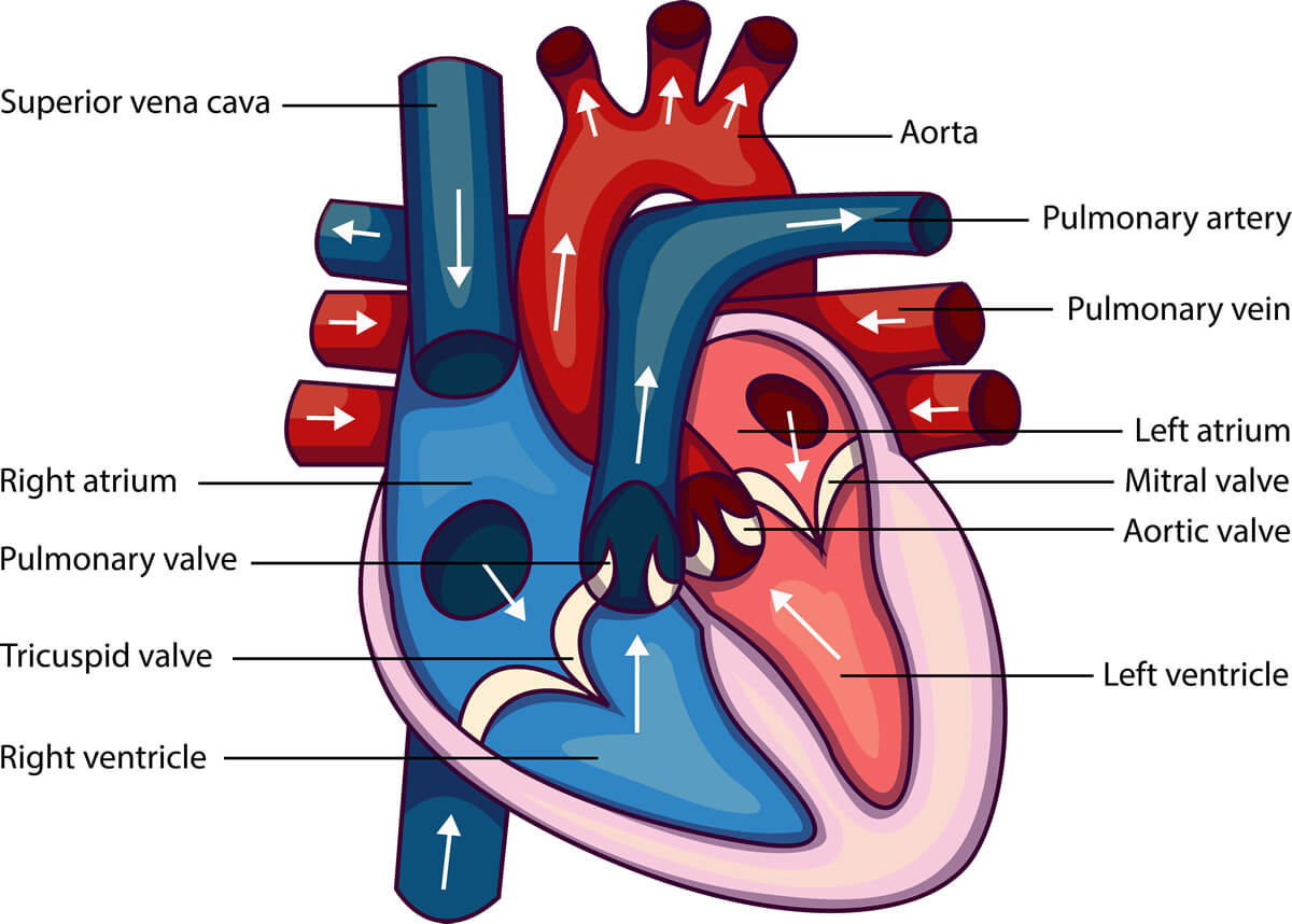
The four-chambered heart is divided into two sides, a right side and a left side, resulting in two distinct halves. Each side of the heart is divided into two chambers: an upper chamber known as the atrium, and a lower chamber known as the ventricle.
An artery transports blood from the heart to the body, whereas a vein transports blood from the body to the heart. It is through the right atrium that venous blood enters the heart, where it travels into the right ventricle. From there it is transported to and expelled from the lungs by way of the pulmonary artery.
The lungs are responsible for removing carbon dioxide from the bloodstream and replacing it with oxygen. The increasingly oxygen-rich blood comes from the lungs and enters the left atrium via the pulmonary vein, where it continues its journey to the left ventricle and beyond.
With the aorta as a conduit, the left ventricle transports oxygenated blood to the rest of the body to nourish the tissues and organs. The pulmonary artery is the only artery in the body that carries deoxygenated blood, and the pulmonary vein is the only vein in the body that carries oxygenated blood. Both are located in the lungs.
Electrical Conduction
The conduction system of your heart is analogous to the electrical wiring of a house. It is in charge of regulating the rhythm and rate of your heartbeat.
It consists of the following:
– the Sinoatrial (SA) node is responsible for sending the impulses that cause your heart to beat
– the atrioventricular (AV) node is responsible for transmitting electrical signals from the upper chambers of your heart to the lower chambers
Your heart is also connected to the outside by an intricate network of electrical bundles and fibers. This network consists of the following:
– the left bundle branch is responsible for sending electrical impulses to your left ventricle
– the right bundle branch is responsible for sending electric impulses to your right ventricle
– the Bundle of His sends impulses from your AV node to the Purkinje fibers
– the Purkinje fibers are responsible for making your heart ventricles contract and pump blood
Blood Circulation
Blood circulation begins when the heart relaxes between two heartbeats. Blood flows from both atria (the top two chambers of the heart) into the ventricles (the lower two chambers of the heart), which then expand as a result of the expansion.
This is followed by the ejection period, during which both ventricles work together to pump blood into the major arteries. The left ventricle is responsible for pumping oxygen-rich blood into the major artery in the systemic circulation (aorta).
As the blood flows from the main artery to the capillary network, it becomes smaller in size as it progresses. There, the blood loses oxygen, nutrition, and other vital substances, while simultaneously gaining carbon dioxide and waste products.
Because the blood is now depleted of oxygen, it collects and flows into veins, where it is transported to the right atrium and into the right ventricle.
This is the point at which the pulmonary circulation begins: the right ventricle pumps low-oxygen blood into the pulmonary artery, which then divides into smaller and smaller arteries and capillaries as it travels down the pulmonary artery.
The capillaries form a tiny network around the pulmonary vesicles (grape-like air sacs at the end of the airways), which helps to protect them from injury. This is the point at which carbon dioxide from the blood is expelled into the air inside the pulmonary vesicles and fresh oxygen is introduced into the bloodstream. Carbon dioxide is expelled from our bodies as we breathe out.
The pulmonary veins and the left atrium carry oxygen-rich blood to the left ventricle, which is located in the heart. The next heartbeat begins a fresh cycle of systemic circulation, which continues until the next heartbeat.
Now you should have a basic yet solid understanding of heart anatomy and function. Take our ACLS course to solidify your learning and go deeper into heart function in the context of providing emergency cardiac assistance. Sign up for our ACLS course today!
